|
Bromodeoxyuridine
(BrdU) Cell Proliferation ELISA Kit
BrdU Proliferation Assay is a
completely HTS compatible, non-isotopic, colorimetric proliferation enzyme-linked
immunosorbent assay (ELISA) assay kit for the detection of bromodeoxyuridine
incorporation into newly synthesized DNA of adherent and non-adherent cells.
Features:
- Colorimetric assay
- HTS compatible format
- Optional spin step
- High sensitivity
- 4 deg C storage with long shelf life
- Non-radioactive
- 2.5 hour protocol
- Suitable for all species
- Suitable adherent or non-adherent cell types
Performance Summary:
- Sensitivity: 40 cells/well
- Intra-assay C.V.'s: <10% (spin protocol)
- Inter-assay C.V.'s: <10% (spin protocol)
Ordering Information:
Cat. No.
X1327K1=
200 tests
X1327K2 = 1000 tests
X1327K3
= 5000 tests
For larger quantity pricing,
please contact Customer Service
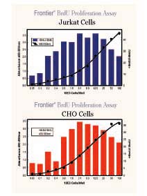
Legend:
BrdU assay. Detection of variable numbers of Jurkat (non-adherent) or CHO cells
(adherent) per well. Y axis-left, OD 450-550nm. Y axis-right, signal-to-noise
ratio.
Protocol Summary: Not to be
used in place of detailed protocol:
1. Cell
Plating – no Test Reagent/Drug
(skip step 3 below) |
• Seed
cells at 1-2 x 10⁵ cells/ml, 100 ml/well |
2. Cell Plating –
withTest Reagent/Drug
(see below step 3) |
• Seed cells at 0.5-4
x 105 cells/ml, 50 ml/well |
| 3. Addition of Test
Reagent(s)/Drug |
• Add 50 ml/well, 2X
concentration desired |
| 4. Addition of BrdU
|
• Dilute 500X stock
BrdU, add 20 ml/well
(be sure to include a No BrdU control) |
| 5. Incubate
|
• 2-24 hours
|
6. Fix
and Denature
- Adherent Cells
|
• Aspirate (or flick) the media from the cell wells
• Add 200 ml/well Fixing Solution
• Incubate 30 minutes at Room Temp.
• Aspirate the Fixing Solution and blot the plates dry. |
- Suspension Cells
No-Spin Procedure |
• Add 200 ml/well
Fixing Solution on top of the cells.
• Incubate 1 hour at Room Temp
• Aspirate the Fixing Solution and blot the plates dry. |
-
Suspension Cells
Spine Procedure |
• Spin the plates for
5 minutes at 1000 rpm.
• Aspirate media, add 200 ml/well Fixing Solution.
• Incubate for 30 minutes, room temp.
• Aspirate the Fixing Solution and blot the plates dry. |
| 7. Wash Step
|
• Wash X3 with 1X
wash buffer and blot dry. |
| 8. Detector Antibody
|
• Add 100 ml/well of
diluted detector antibody. |
| 9. Incubate
|
• 1 hour at room temp. |
| 10. Wash Step
|
• Wash X3 with 1X
wash buffer and blot dry. |
| 11. Conjugate
Addition |
• Add 100 ml/well HRP-conjugate |
| 12. Incubate
|
• Incubate for 30
minutes at room temperature. |
| 13. Wash Step and
Final Water Wash |
• Wash as above.
Perform a final distilled water wash by flooding the entire plate
with distilled water. Pat dry on absorbent paper towels. |
14.
Development
|
• Add 100
ml/well TMB Peroxidase substrate |
| 15.
Incubate |
• 30
minutes at room temperature in the dark. |
| 16. Stop |
• Add 100 ml of acid
Stop Solution to every well |
| 17. Read |
• Read the at 450/550
nm |
| 